
A recent paper in The Lancet Digital Health shows that the brain can act in a surprising way after a stroke. Scientists at USC’s Stevens Institute discovered that people with serious movement problems after a stroke often have a "younger" looking brain in parts that were not damaged.
The team studied brain scans from more than 500 stroke survivors collected at 34 centers in eight countries. They used advanced computer models that had learned from tens of thousands of MRI scans to estimate the "brain age" of many regions in each hemisphere.
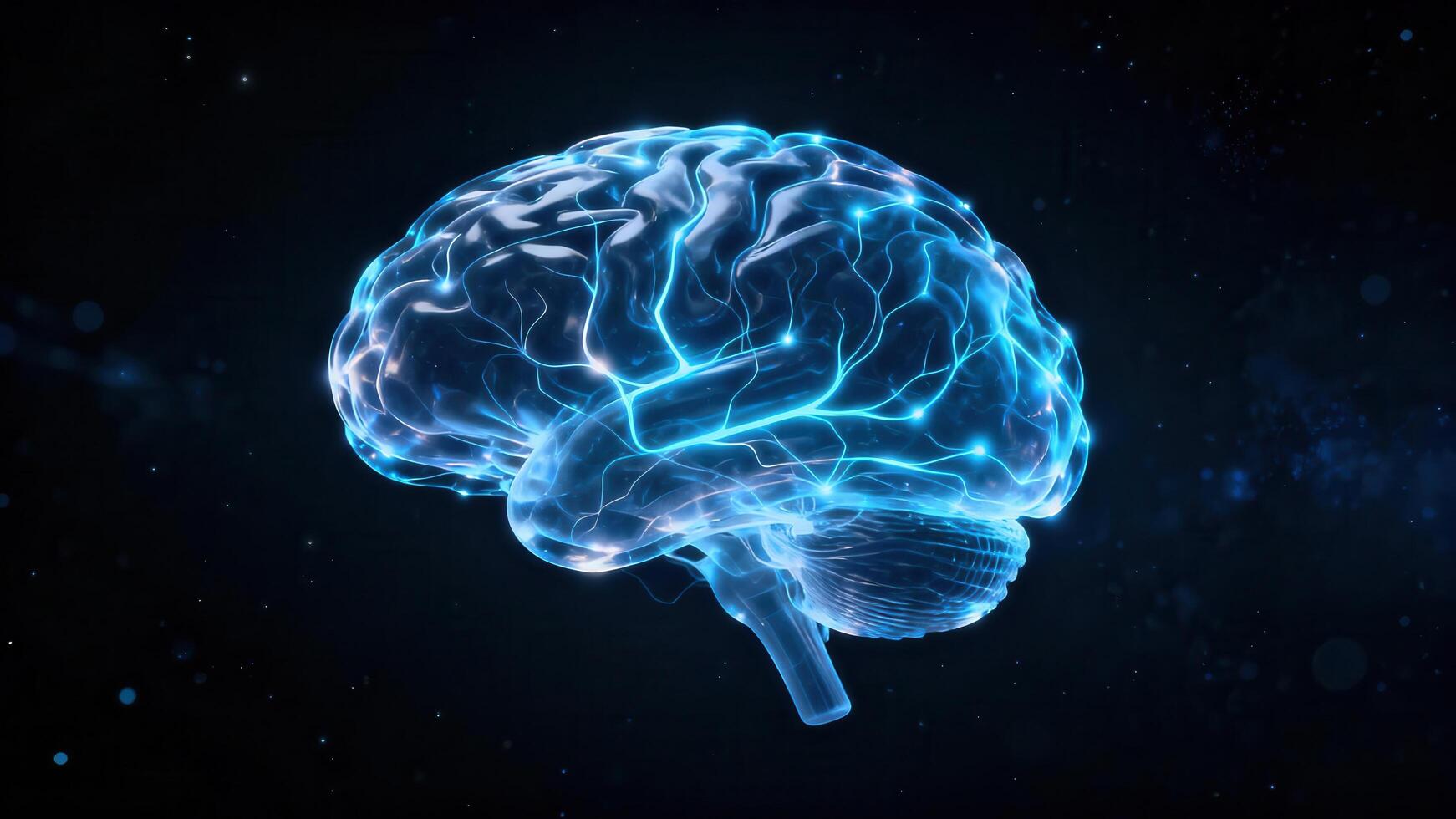
AI Shows Brain Rewiring
To do this, researchers used a type of artificial intelligence called a graph convolutional network. The program guessed the biological age of 18 brain regions from the MRI data. By comparing this guess with the person’s real age, they got a measure called brain‑predicted age difference (brain‑PAD), which tells how healthy the brain looks.
When they matched brain‑PAD scores with motor‑function tests, a clear pattern appeared. Stroke survivors who still had strong movement loss after more than six months of therapy showed a younger‑than‑expected brain age in the opposite side of the brain. The effect was strongest in the frontoparietal network, a region that helps plan movement, pay attention, and coordinate actions.
"When stroke damage causes big movement loss, the healthy side of the brain may try to help," said lead researcher Kim. "We saw this in the opposite frontoparietal network, which looked more youthful. It’s not a sign that movement is fully back, but rather that the brain is trying to adjust when the damaged side can’t work properly. This gives us a new view of neuroplasticity that regular scans miss."
Big Data Reveals Hidden Patterns
The work relied on ENIGMA, a worldwide partnership that gathers brain data from over 50 countries. By standardizing MRI and clinical information from many groups, the team created the largest stroke‑imaging dataset ever.
"By combining data from hundreds of stroke patients worldwide and using cutting‑edge AI, we can spot tiny patterns of brain re‑organization that smaller studies would miss. These findings could eventually help doctors design personalized rehab plans," said Arthur W. Toga, PhD, director of the Stevens Institute.
Moving Toward Personalized Stroke Care
The researchers will keep following patients from the early weeks after a stroke through long‑term recovery. Watching how brain‑age patterns change over time could help doctors match treatments to each person’s unique healing process, aiming for better outcomes and quality of life.
The study, titled "Deep learning prediction of MRI‑based regional brain age reveals contralesional neuroplasticity associated with severe motor impairment in chronic stroke: A worldwide ENIGMA study," was funded by the National Institutes of Health (NIH) grant R01 NS115845 and involved collaborators from institutions such as the University of British Columbia, Monash University, Emory University, and the University of Oslo.