
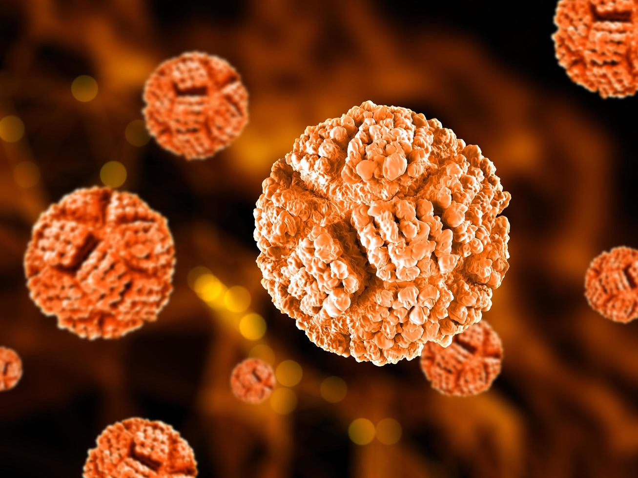
Researchers at the University of Maryland, Baltimore County have found a key step that enteroviruses use to make copies of themselves inside human cells. The work, published in Nature Communications, explains how viruses that cause polio, encephalitis, heart inflammation, and even the common cold take over the cell’s machinery.
The study was led by Deepak Koirala, an associate professor of chemistry and biochemistry, together with recent Ph.D. graduate Naba Krishna Das. Their experiments answer long‑standing questions about how these tiny RNA viruses begin to replicate after they enter a cell.
How Enteroviruses Copy Their Genetic Material
Enteroviruses carry a very small RNA genome. This RNA must do two jobs at once: direct the production of viral proteins and serve as a template for making new viral RNA.
Most of the genome codes for the virus’s outer shell proteins. It also contains instructions for a special fusion protein called 3CD. The “3C” part cuts long protein chains into the pieces the virus needs, while the “3D” part works as an RNA polymerase that copies the viral RNA. Human cells do not have this polymerase, so the virus brings its own.
Using X‑ray crystallography, the team captured the shape of a cloverleaf‑shaped region of viral RNA together with the 3CD protein. They discovered that the 3C portion of 3CD grabs onto the cloverleaf and then pulls in a host protein named PCBP2. This creates a replication complex that can start copying the virus’s RNA.
The complex acts like a switch. When 3CD is attached, the virus makes copies of its RNA. When it detaches, the RNA is free to be read as a message for making viral proteins.
Clearing Up a Scientific Puzzle
The researchers used several techniques—X‑ray crystallography, isothermal titration calorimetry, and biolayer interferometry—to watch how the molecules interact. Their data showed that two full 3CD molecules bind side‑by‑side on the cloverleaf RNA. Earlier studies had suggested a single fused pair, so this finding resolves a long‑standing debate.
Why the virus needs two copies of 3CD is still unclear, but the new picture clarifies the first steps of viral replication.
Opportunities for New Antiviral Medicines
The team examined seven different enteroviruses and found that all of them use almost the same cloverleaf shape and binding pattern. This similarity means the structure is essential for the virus to survive. Changing it would likely stop the virus from replicating.
Because the RNA cloverleaf is so conserved, it could become a target for broad‑spectrum antiviral drugs that work against many enteroviruses at once, rather than a single strain.
Scientists are already creating drugs that block the 3C and 3D parts of the protein. The new study suggests another strategy: designing molecules that disrupt the RNA‑protein interaction itself.
“Now we can design drugs that fit exactly into the interface we just mapped,” Koirala said. “Targeting the RNA or the spot where it meets the protein could be a powerful new approach.”
The research highlights how clever viruses can be, even with genomes the size of a single human messenger RNA. Understanding these basic mechanisms is the first step toward turning them into drug targets that could protect people from many serious illnesses.